INTRODUCTION
Because
of their extreme minuteness, bacteria are not generally studied with the low
power or high power dry objectives. Instead they are stained and observed with
the oil immersion objective.
The wet
mount method enables you to study the sizes and shapes of living
microorganisms. It is also enables you to determine if cells are motile. The
wet mount method is quick and easy, and does not require special equipment.
OBJECTIVE
~ To provide an
experience in the use of microscope
~ To illustrate the diversity of cells and microorganisms
RESULT

Typical bacillus
40x magnification

Typical bacillus
100x magnification

Typical bacillus
400x magnification

Lactobacillus fermentum
1000x magnification

Lactobacillus fermentum
400x magnification
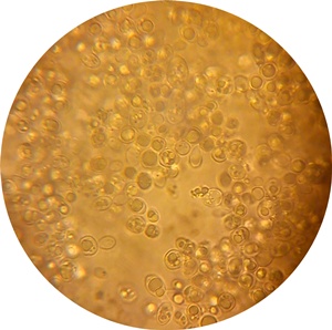
Sacchamnyces arerisiae
1000x magnification

Sacchamnyces arerisiae
400x magnification
DISCUSSION
1. Stained cells
By using technique called the Gram stain, we can classify it is Gram
positive bacteria. It is rod shape and typical bacillus. Gram positive bacteria
has a thick cell wall made of peptidoglycan that traps crystal violet in the
cytoplasm. The masks the added red safranin dye. Most bacterial cell walls
contain peptidoglycan, a network of modified-sugar polymers cross-linked by
short polypeptides. So when we observe under microscope we use different
magnification to get better view which is 4x, 10x and 40x.
2. Wet mount
Second experiment we use wet
mount methods which helps us to study the sizes and shapes of living organisms.
In wet mount, the specimen suspended in drop of liquid method, it is quick
preparation and it is possible to observe living and moving organisms. We also
observe the organisms using different magnification which is 40x,100x,400x and
1000x magnification. Immersion of oil is used to observed under 1000x
magnification to get clearer view and brightness. This characteristics are most
critical under higher magnification.
We also practice
aseptic technique during the experiment. We should slowly heat the inoculating
wire until it glow orange. Then, cool it before attempting any transfer of
material. We also heat the mouth of an opening bottle. This is to make sure we
are not get infection and prevent spread of microorganisms.
During this
experiment, we observe Lactobacillus fermentum under 400x and 1000x
magnification. Thus we can see s rod-shaped structure and also the organisms is
moving. Lactobacillus cannot be seen under the 40x and 100x magnification
because it is too tiny to be seen. Same goes to yeast, we unable to see it
under 40x and 100x magnification but can be seen under 400x and 1000x
magnification. Yeast and fungi which do not contain chlorophyll. Under
microscope we can see it is cocci or spherical shape. We can see clearer image
of shape when use oil immersion at 1000x magnification.
CONCLUSION
Gram-positive bacteria ang Gram-negative bacteria. These staining reaction take advantages of the facts that the cells or structure within the cell display dissimilar staining reactions that can be distinguished by the used of different dyes. A gram positive bacteria can be identified by retaining a purple colour dye within its peptidoglycan exterior. This staining technique is used to help identify various bacteria. The gram positive bacteria that are purple hold stain due to it's layered cell membrane. It contain a peptidoglycan layer acts as a lattice trapping the crystal violet-iodine dye complex. Typical bacillus has a rod-shaped and the average bacillus is 0.5-1.0 um wide by 1.0-4.0 um long.
Many sample look better when use immersion oil. This is known as a wet mount. Oil helps support the sample and it fills the space between the cover slip and the slide allowing light to pass easily through the slide, the sample and cover slip. Lactobacillus fermentum has been identified as potential probiotic. The use of gut microbes as probiotics in food is aim towards preventing and treating various health disease. We also use wet mount method to see yeast clearly under microscope using 400x and 1000x magnification.
REFERENCES
1. Pearson International Biology, 8th Edition, Campbell,Reece
2.http://en.wikipedia.org/wiki/Bacillus_(shape)
3.http://www.nutritionj.com/content/10/1/30
4. http://www.ncbi.nlm.nih.gov/pmc/articles/PMC119863/
No comments:
Post a Comment